A neuron is an electrically excitable cell that receives and transmits informations through synapses. Neurons are the cells that compose the central nervous system, which includes the brain and the spinal cord, and the peripheral nervous system, which comprises somatic nervous system and the autonomic nervous system.
1) The structure of a neuron
Neurons are composed by a cell body or soma from where comes an axon and an arborization composed by dendrites. Neurons have many dendrites, but only one axon.
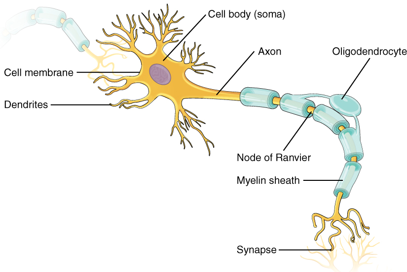
The cell body contains the usual organelles that the other cells contain too: nucleus, endoplasmic reticulum, golgi apparatus, nucleolus, mitochondria, ribosomes, endosomes, ribosomes and peroxisomes. Some of these cell elements act for the expression of the genetic information and for the synthesis of proteins for energy production and growth for example.

The dendrites receive the influxes coming from another neuron, and more precisely from axons of another neuron. The dendritic arborization may branch very extensively and it then forms what is called dendritic spines.
Indeed, axons transmit the influx coming from the cell body. Axons’ length can vary from only one millimeter to one meter, like the ones going from the brain to the spinal cord. The axon terminal gives rise to a synapse where chemical messengers called neurotransmitters are released when the influx reaches this region. The transmitter neuron is called the presynaptic neuron and the neuron which receives the information is called the postsynaptic neuron. We will develop these elements and the synapse in the second part.
Some axons are surrounded by a myelin sheath, which is a layer of fatty acids that allows the electrical message to be spread more quickly. In the brain and spinal cord, the myelin sheath is formed from cells called oligodendrocytes that wrap their extensions around axons. In nerves outside spinal cord, the schwann cells produce the myelin sheath. As the myelin sheath is discontinuous, there are myelin-sheath gaps that are called node of Ranvier which are highly enriched with ions. When a nerve impulse is spread along an axon, it “jumps” from a node of Ranvier to a node of Ranvier; this action is called the saltatory conduction.
Finally, the region in the cell body where the axon originates is called the axon hillock. There is not any ribosomes and most of other organelles in the axon hillock, and the neurofilaments (a type of intermediate filaments that maintain neuron’s shape and mechanical strength) are grouped as a fascicle. There are however some organelles that are present in the axon hillock which are then transported down the axon. The initial segment is the region between the axon hillock and the beginning of the myelin sheath.
In Figure 1.3 you can see a comparison of a nervous message spreading (from the cell body to a synapse) between a myelinated axon (the right one) and an unmyelinated axon (the left one).

2) The neuron’s membrane : membrane potential, ion channels
Now that we have talked about the structure of the neuron, we can go deeper into its description, and more precisely into the description of its membrane (5nm thickness).
First, let’s deal with the membrane potential, or resting membrane potential if the neuron is at rest; it is generally about -60mV and can go until -80mV and it is an electrical potential difference.
Where does this difference comes from? It is related to the difference of ions between the intracellular and extracellular space: the intracellular space has a more negative charge than the extracellular space, due to the negatively charged proteins and phosphate ions inside the neuron.
Indeed, in neurons, the most three abundant ions are chloride (Cl-), sodium (Na+) and potassium (K+). Inside neurons, there are mostly potassium ions and outside neurons there are mostly sodium ions and ions chloride.
Here are the following concentrations :
| Concentration in mM intracellular space | Concentration in mM extracellular space | |
| Sodium ions Na+ | 15 | 150 |
| Ions chloride Cl– | 10 | 120 |
| Potassium ions K+ | 140 | 5 |
How do the ions flow from the intracellular space to the extracellular space and vice-versa? This is through selective ions channels : sodium channels, potassium channels, and chloride channels in neurons, and through sodium-potassium pumps.
These ion channels are also called passive channels (or leakage channels) and they are all located in the cell membrane all over the neuron.
As the ions’ concentrations are different inside and outside the cell, there are concentration gradients for each ions. But there is also an electrical gradient due to the charge difference between the extracellular space and intracellular space.
There is indeed a diffusion through the selective ion channels, but there are very few sodium ions channels open whereas there are a lot of potassium ion channels that are open so a lot of potassium ions can flow outside the cell. As the potassium ions positively charged leave the intracellular space (more than the sodium ions enter the intracellular space), it lets a negative charge inside the neuron. Also, the sodium-potassium pump pumps three sodium ions out of the cell and two potassium ions inside the cell, against the concentration gradient (using ATP): it leads to the creation and the maintaining of the membrane potential.
Then, when the neuron is at equilibrium, the electrical gradient balances exactly the concentration gradient, so there won’t be any flow for a specific ion.
The equilibrium (or reversal) potential for a specific ion is the electrical potential where chemical and electrical potentials are in balance. We can actually calculate the equilibrium potential of the ions we have talked above, with the Nernst equation:

– E in mV
– R = 8,314 J/mol/K
– z charge of the ion
– T temperature in K
– F = 96485 A/mol
Here are the equilibrium potential of the main ions of neurons at 37°C:
| Equilibrium potential in mV | |
| Sodium ions Na+ | +60 |
| Ions chloride Cl– | -70 |
| Potassium ions K+ | -90 |
3) Types of neurons
Neurons can be classified into four different groups according to their structure:
– Unipolar neurons: the simplest category of neurons. They have a single extension that gives rise to branches, and some of them are dendrites.
– Bipolar neurons: they have an axon (that transmits signals from the cell body to the brain and spinal cord) and dendrites (that send signals from the organs of the body to the cell body). They carry the light signals produced by the photoreceptors in the outer retina to amacrine cells and ganglion cells in the inner retina.
– Pseudounipolar neurons: they are variants of bipolar neurons. The axon attached to the cell body is linked to two opposite poles (one towards the muscle and skin and the other towards the spinal cord).
– Multipolar neurons: these are the common model neurons we usually see in neuron structure diagrams. They have a cell body from where an axon and dendrites come from.

It is also possible to split the neurons into three different types according to their function: sensory neurons, motor neurons and interneurons.
First, sensory neurons are the nerve cells which are activated by a sensory input coming from the environment. These inputs can be light, sound, touch, heat, taste, smell. They send the signals to the rest of the nervous system from the information they received. Most of them are pseudo-unipolar.
Then, motor neurons transmit signals from the brain to the spinal cord and then to the muscle cells. They are multipolar.
Finally, interneurons connect the spinal cord and sensory neurons. They transfer signals between sensory and motor neurons, but they can also communicate with each other. They are multipolar.
4) The support cells : glia
We have seen the structure of a neuron and the function of its components, the ions that produce the electrical potential difference, and the different types of neurons according to their function or to their structure. However neurons cannot stay “assembled” and maintain their shape just like that. They need other cells, called glia (or glial cells or neuroglia) which are non-neuronal cells of the central nervous system and peripheral nervous system.
There is a big variety of glial cells, each having a particular function, and some are present in the central nervous system and others in the peripheral nervous system. Here are all the different types of glial cells:
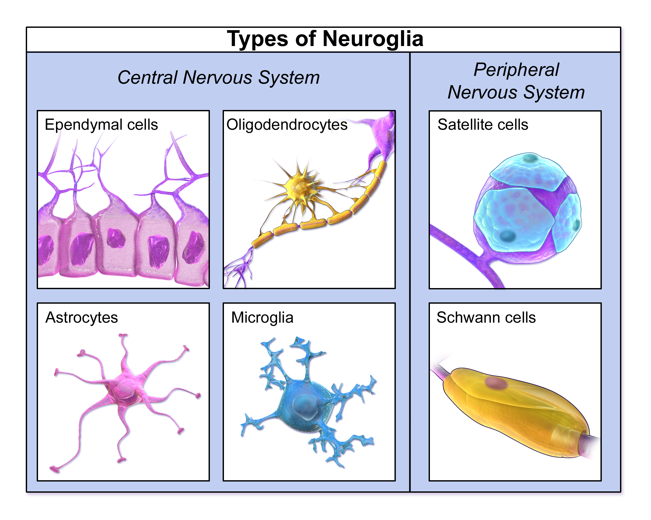
We will just develop on the main glial cells of the central nervous system.
a) Microglia
Microglia represents the cells of the immune system of the brain, which protect the brain against injuries and diseases from pathogenic factors. In the central nervous system, under physiological conditions, microglia exists in what we call their “resting state”: they have a small cell body and thin protuberances that extend branches in all directions. At this resting state, they are constantly moving in their own territory. When an insult is detected in the brain, the microglia leaves its resting state, by a process called microglial activation. During this process, its cell body is enlarged and its protuberances become thicker and fewer, and it begins to produce immune response molecules. Then some microglia return in proliferative state and some other multiply on the contaminated region. Sometimes, when the brain cells are still destroyed by the pathogen and the infection persists, microglia can turn into phagocytes.
b) Astrocytes
Astrocytes are the most numerous glial cells in the central nervous system. They have the shape of a star and maintain the neuron’s working environment by controlling the levels of neurotransmitters around the synapses and the concentrations of some ions. They have very heterogeneous morphologies, they can be classified into two main groups: protoplasmic astrocytes in gray matter of the brain, and fibrous astrocytes in white matter of the brain. Protoplasmic astrocytes have thin and complex processes (approximately 50 micrometers long) and form many contacts with neurons. Fibrous astrocytes have very long processes (up to 300 micrometers long) but less elaborate than protoplasmic astrocytes.
An area of research suggests that astrocytes also play a role in modulating how neurons communicate.
c) Oligodendrocytes
The main functions of oligodendrocytes are the production of myelin around axons and the support to axons in the central nervous system.
Then oligodendrocytes are mostly present in white matter, but also in gray matter. These oligodendrocytes of gray matter are called satellite oligodendrocytes as they are not directly linked to myelin sheath, but their function is still not really well known.
During the development, there are first oligodendrocytes precursors found in other parts of the brain, that then migrate to their destination where they differentiate into mature oligodendrocytes.
After that, they can play their role (for example) of myelin producer around axons.
