Have you ever asked yourself how the information is transmitted from our brain to our body into an action ? All our actions are responses to stimulation coming from our environment.An example of one of these reactions can be illustrated by the reflex of taking back your hand when touching a hot glass without any thoughts behind it .
For decades the Egyptians thought that the seat of intelligence was the heart. The first people that studied the brain were the Greeks who attempted to know what was the role of brain and how it worked, to explain some brain disorders.
The main discoveries in neurology were during the 18th century when Luigi Galvani explained in 1791 the electricity in nerves. After,J. E. Purkinje came with the first description of a neuron. In the 19th century, shortly after J. E. Purkinje, the doctor and scientist Paul Broca discovered that the different regions of the brain were dedicated to a function, his main discovery was the center of speech in the brain that today is known as the Broca’s area (look at chapter 49 Nervous system). However, at this time the relationship between nerve cells, axons, and dendrites were not really well-known and clear, until Santiago Ramon y Cajal introduced the neuron doctrine as we use till today, saying that neurons are independent cell units which make up the nervous system.
The 20th century has been a revolution to study the brain, the neurons, the synapses or the signal transmission thanks to all the new techniques such as the Magnetic Resonance Imaging (MRI) invented in the 1970s, the electrophysiology from the 1950s (to study the electrical properties of living neurons), or the confocal microscopy described in 1953 and available around 1980.
At the end of this chapter you should know the structure of a neuron, the environment of neurons, how neurons communicate with each other, how the electrical signal is transmitted from a neuron to another, and we will also present some diseases related to neuronal dysfunctions.
A neuron is an electrically excitable cell that receives and transmits informations through synapses. Neurons are the cells that compose the central nervous system, which includes the brain and the spinal cord, and the peripheral nervous system, which comprises somatic nervous system and the autonomic nervous system.
1) The structure of a neuron
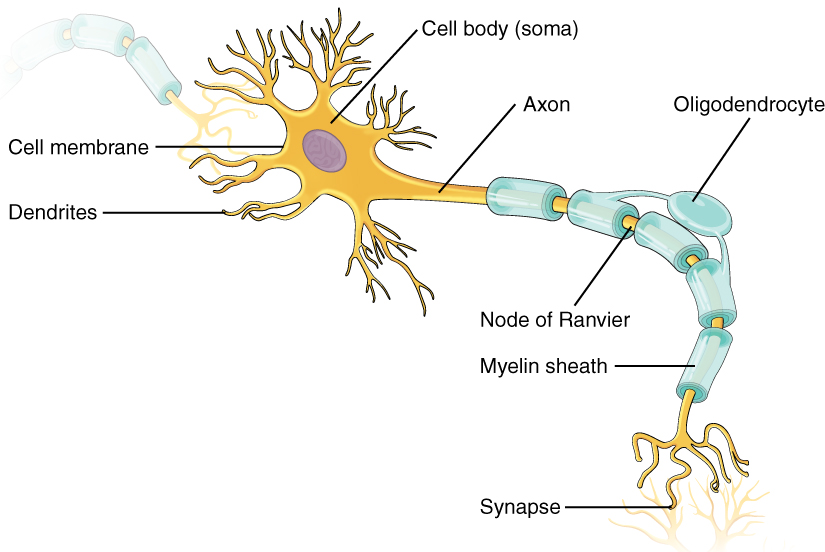
Neurons are composed by a cell body or soma from where comes an axon and an arborization composed by dendrites. Neurons have many dendrites, but only one axon.
Figure 1.1: model structure of a neuron
The cell body contains the usual organelles that the other cells contain too: nucleus, endoplasmic reticulum, golgi apparatus, nucleolus, mitochondria, ribosomes, endosomes, ribosomes and peroxisomes. Some of these cell elements act for the expression of the genetic information and for the synthesis of proteins for energy production and growth for example.
Figure 1.2: detailed elements inside a neuronal cell body
The dendrites receive the influxes coming from another neuron, and more precisely from axons of another neuron. The dendritic arborization may branch very extensively and it then forms what is called dendritic spines.
Indeed, axons transmit the influx coming from the cell body. Axons’ length can vary from only one millimeter to one meter, like the ones going from the brain to the spinal cord. The axon terminal gives rise to a synapse where chemical messengers called neurotransmitters are released when the influx reaches this region. The transmitter neuron is called the presynaptic neuron and the neuron which receives the information is called the postsynaptic neuron. We will develop these elements and the synapse in the second part.
Some axons are surrounded by a myelin sheath, which is a layer of fatty acids that allows the electrical message to be spread more quickly. In the brain and spinal cord, the myelin sheath is formed from cells called oligodendrocytes that wrap their extensions around axons. In nerves outside spinal cord, the schwann cells produce the myelin sheath. As the myelin sheath is discontinuous, there are myelin-sheath gaps that are called node of Ranvier which are highly enriched with ions. When a nerve impulse is spread along an axon, it “jumps” from a node of Ranvier to a node of Ranvier; this action is called the saltatory conduction.
Finally, the region in the cell body where the axon originates is called the axon hillock. There is not any ribosomes and most of other organelles in the axon hillock, and the neurofilaments (a type of intermediate filaments that maintain neuron’s shape and mechanical strength) are grouped as a fascicle. There are however some organelles that are present in the axon hillock which are then transported down the axon. The initial segment is the region between the axon hillock and the beginning of the myelin sheath. In Figure 1.3 you can see a comparison of a nervous message spreading (from the cell body to a synapse) between a myelinated axon (the right one) and an unmyelinated axon (the left one).
Figure 1.3: spreading of an electrical signal in an unmyelinated axon (left) and in a myelinated axon (right)
2) The neuron’s membrane : membrane potential, ion channels
Now that we have talked about the structure of the neuron, we can go deeper into its description, and more precisely into the description of its membrane (5nm thickness).
First, let’s deal with the membrane potential, or resting membrane potential if the neuron is at rest; it is generally about -60mV and can go until -80mV and it is an electrical potential difference.
Where does this difference comes from? It is related to the difference of ions between the intracellular and extracellular space: the intracellular space has a more negative charge than the extracellular space, due to the negatively charged proteins and phosphate ions inside the neuron. Indeed, in neurons, the most three abundant ions are chloride (Cl-), sodium (Na+) and potassium (K+). Inside neurons, there are mostly potassium ions and outside neurons there are mostly sodium ions and ions chloride. Here are the following concentrations :
Concentration in mM intracellular space
Concentration in mM extracellular space
Sodium ions Na+
15
150
Ions chloride Cl–
10
120
Potassium ions K+
140
5
How do the ions flow from the intracellular space to the extracellular space and vice-versa? This is through selective ions channels : sodium channels, potassium channels, and chloride channels in neurons, and through sodium-potassium pumps. These ion channels are also called passive channels (or leakage channels) and they are all located in the cell membrane all over the neuron.
As the ions’ concentrations are different inside and outside the cell, there are concentration gradients for each ions. But there is also an electrical gradient due to the charge difference between the extracellular space and intracellular space. There is indeed a diffusion through the selective ion channels, but there are very few sodium ions channels open whereas there are a lot of potassium ion channels that are open so a lot of potassium ions can flow outside the cell. As the potassium ions positively charged leave the intracellular space (more than the sodium ions enter the intracellular space), it lets a negative charge inside the neuron. Also, the sodium-potassium pump pumps three sodium ions out of the cell and two potassium ions inside the cell, against the concentration gradient (using ATP): it leads to the creation and the maintaining of the membrane potential.
Then, when the neuron is at equilibrium, the electrical gradient balances exactly the concentration gradient, so there won’t be any flow for a specific ion. The equilibrium (or reversal) potential for a specific ion is the electrical potential where chemical and electrical potentials are in balance. We can actually calculate the equilibrium potential of the ions we have talked above, with the Nernst equation:
– E in mV – R = 8,314 J/mol/K – z charge of the ion – T temperature in K – F = 96485 A/mol
Here are the equilibrium potential of the main ions of neurons at 37°C:
Equilibrium potential in mV
Sodium ions Na+
+60
Ions chloride Cl–
-70
Potassium ions K+
-90
3) Types of neurons
Neurons can be classified into four different groups according to their structure:
– Unipolar neurons: the simplest category of neurons. They have a single extension that gives rise to branches, and some of them are dendrites. – Bipolar neurons: they have an axon (that transmits signals from the cell body to the brain and spinal cord) and dendrites (that send signals from the organs of the body to the cell body). They carry the light signals produced by the photoreceptors in the outer retina to amacrine cells and ganglion cells in the inner retina. – Pseudounipolar neurons: they are variants of bipolar neurons. The axon attached to the cell body is linked to two opposite poles (one towards the muscle and skin and the other towards the spinal cord). – Multipolar neurons: these are the common model neurons we usually see in neuron structure diagrams. They have a cell body from where an axon and dendrites come from.
Figure 1.4: different types of neurons according to their structure
It is also possible to split the neurons into three different types according to their function: sensory neurons, motor neurons and interneurons. First, sensory neurons are the nerve cells which are activated by a sensory input coming from the environment. These inputs can be light, sound, touch, heat, taste, smell. They send the signals to the rest of the nervous system from the information they received. Most of them are pseudo-unipolar. Then, motor neurons transmit signals from the brain to the spinal cord and then to the muscle cells. They are multipolar. Finally, interneurons connect the spinal cord and sensory neurons. They transfer signals between sensory and motor neurons, but they can also communicate with each other. They are multipolar.
4) The support cells : glia
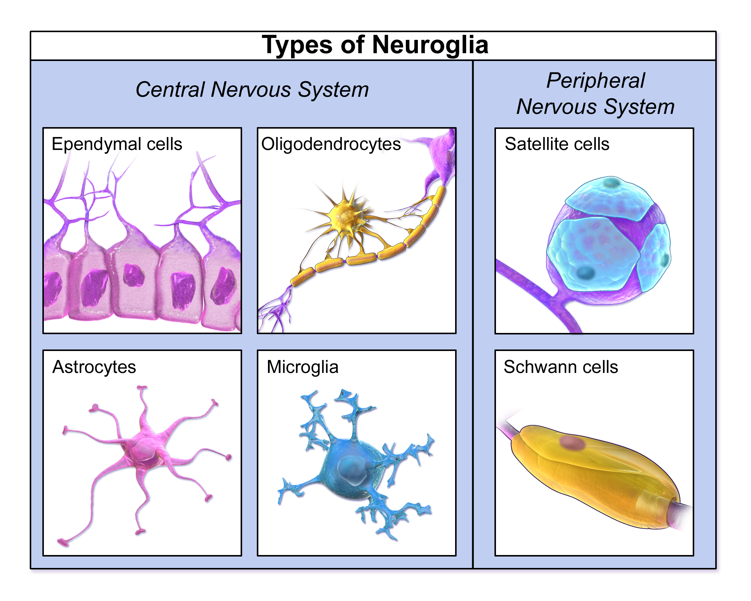
We have seen the structure of a neuron and the function of its components, the ions that produce the electrical potential difference, and the different types of neurons according to their function or to their structure. However neurons cannot stay “assembled” and maintain their shape just like that. They need other cells, called glia (or glial cells or neuroglia) which are non-neuronal cells of the central nervous system and peripheral nervous system. There is a big variety of glial cells, each having a particular function, and some are present in the central nervous system and others in the peripheral nervous system. Here are all the different types of glial cells:
Figure 1.5: different types of glial cells in the central nervous system and in the peripheral nervous system
We will just develop on the main glial cells of the central nervous system.
a) Microglia Microglia represents the cells of the immune system of the brain, which protect the brain against injuries and diseases from pathogenic factors. In the central nervous system, under physiological conditions, microglia exists in what we call their “resting state”: they have a small cell body and thin protuberances that extend branches in all directions. At this resting state, they are constantly moving in their own territory. When an insult is detected in the brain, the microglia leaves its resting state, by a process called microglial activation. During this process, its cell body is enlarged and its protuberances become thicker and fewer, and it begins to produce immune response molecules. Then some microglia return in proliferative state and some other multiply on the contaminated region. Sometimes, when the brain cells are still destroyed by the pathogen and the infection persists, microglia can turn into phagocytes.
b) Astrocytes Astrocytes are the most numerous glial cells in the central nervous system. They have the shape of a star and maintain the neuron’s working environment by controlling the levels of neurotransmitters around the synapses and the concentrations of some ions. They have very heterogeneous morphologies, they can be classified into two main groups: protoplasmic astrocytes in gray matter of the brain, and fibrous astrocytes in white matter of the brain. Protoplasmic astrocytes have thin and complex processes (approximately 50 micrometers long) and form many contacts with neurons. Fibrous astrocytes have very long processes (up to 300 micrometers long) but less elaborate than protoplasmic astrocytes. An area of research suggests that astrocytes also play a role in modulating how neurons communicate.
c) Oligodendrocytes The main functions of oligodendrocytes are the production of myelin around axons and the support to axons in the central nervous system. Then oligodendrocytes are mostly present in white matter, but also in gray matter. These oligodendrocytes of gray matter are called satellite oligodendrocytes as they are not directly linked to myelin sheath, but their function is still not really well known. During the development, there are first oligodendrocytes precursors found in other parts of the brain, that then migrate to their destination where they differentiate into mature oligodendrocytes. After that, they can play their role (for example) of myelin producer around axons.
Now that you have learned all about a single neuron, imagine a community of neurons. Individual neurons make connections to target neurons and stimulate or inhibit their activity, forming circuits that can process incoming information and carry out a response. In this part you will learn about how neurons communicate and send signals in order to have an action or a response.
1) Signal transmission
Neurons communicate with one another at junctions called synapses. Synapses are usually between an axon and a dendrite of another neuron but there are different types of synapses as you can see in the figure 2.1. In a neuron synapse we can find the presynaptic, or sending neuron, that causes the transmission of a signal from axon terminal to dendrite of another neuron, the postsynaptic, or receiving neuron.
Figure 2.1: different types of synapses
Depending on the type of the stimulator, we can define two types of synapses: electrical and chemical synapses.
a) Chemical synapses
Chemical transmission involves a release of chemical messengers known as neurotransmitters. Neurotransmitters carry information from the presynaptic neuron to the postsynaptic neuron. Inside the axon terminal of a sending cell, there are many synaptic vesicles. These are membrane-bound spheres filled with neurotransmitter molecules. There is a small gap between the axon terminal of the presynaptic neuron and the membrane of the postsynaptic cell, and this gap is called the synaptic cleft.
Now that you know the definitions let’s see how the signal is transmitted in a chemical synapse.
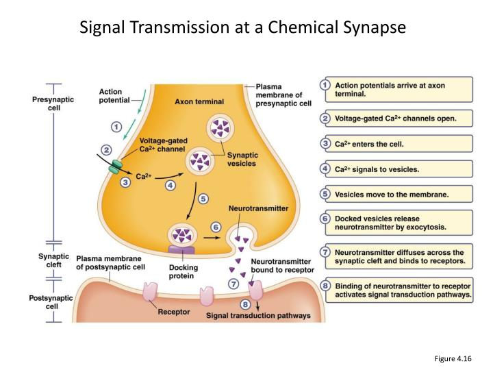
First, the action potential or nerve impulse arrives at the axon terminal (synaptic terminal) of a presynaptic neuron. The primary function of the synaptic terminal is to propagate action potentials from the axon to the presynaptic membrane. Recall that propagation of an action potential requires the presence of both voltage-gated Na+ and K+ channels. The depolarization of the neuron will activate the voltage-gated calcium channels in the cell membrane. The electrical and chemical gradients for Ca2+ ions leads the Ca2+ ions to flow inside the neuron (calcium is present at a much higher concentration outside the neuron than inside). Therefore the calcium rushes into the cell. The Ca2+ allows synaptic vesicles to fuse with the docking protein at the axon terminal membrane and to release the neurotransmitters into the synaptic cleft by exocytosis. Neurotransmitters then diffuse across the synaptic-cleft and bind to the receptors or ligand-gated channels of the postsynaptic cell. The activation of this membrane receptors leads to a change in opening or closing ion membrane channels. These effects on channels behavior can be direct when the receptor is a ligand-gated ion channel, or in other cases, the receptor is not an ion channel itself but activates ion channels through a signaling pathway. This effect in behavior of channels can be in two types depending on the ions involved: –Depolarization makes interior of the cell more positive. –Hyperpolarization makes interior of the cell more negative than its resting value.
Figure 2.2: signal transmission at a chemical synapse
After the release of the neurotransmitters in the synaptic cleft, they fix themselves on the receptors of the postsynaptic neuron, as we said above. However, the neurotransmitters need to be deactivated or removed. If not, there is a continuous activation and action potential firing on the postsynaptic neuron. There are two main mechanisms of neurotransmitter deactivation: enzyme degradation and reuptake.
— The first mechanism, enzyme degradation can be typically applied to the neurotransmitter acetylcholine. When acetylcholine binds to the postsynaptic neuron receptor and depolarizes the cell (this mechanism will be explained later in part 2.2A of this chapter), it quickly unbinds the receptors to bind to the enzyme acetylcholinesterase. This enzyme dissociates the acetate and choline that form acetylcholine by breaking the ester bond which joins these two compounds. Acetylcholine is thus deactivated and cannot bind anymore to the receptors of the postsynaptic neuron.
— The second mechanism could be assimilated to a recycling process: the neurotransmitters are removed from the synaptic cleft with transporter proteins that take them back to the presynaptic terminal where they are packaged again in the vesicles. One of these neurotransmitters that are recycled in this way is serotonin. There is indeed the serotonin transporter called SERT which is a sodium, potassium and chloride dependent receptor. The SERT first binds a sodium ion. Then, serotonin under its protonated form (5HT+) binds to the SERT and finally a chloride ions binds too. It leads to a change in the conformation of the transporter protein, which can cross the membrane of the presynaptic neuron to release the ions and the serotonin in the intracellular space. It then binds to a potassium ion that allows it to go out back in the extracellular space.
b) Electrical synapses
In electrical synapses, unlike chemical synapses, the two neurons are in physical connection. This connection between the presynaptic neuron and postsynaptic neuron is in a form of channel called gap junction, that allows the current-ions flow direct throw them. In an electrical synapse the two cells can exchange cytoplasm therefore the transmission of signals is quicker, and can occur in both directions. This advantage is especially obvious in neuronal pathways that serve as escape mechanisms, such as those in the tail muscles of crayfish and lobsters and those in the pectoral fins of fishes. In these cases, the rapidity of the animal’s response to imminent danger has a selective value. Also, electrical synapses contribute to the synchronized activity of groups of cells. In many cases, they can carry current in both directions so that the depolarization of a postsynaptic neuron will lead to the depolarization of a presynaptic neuron.
Figure 2.3: electrical synapse
Some synapses can be both electrical and chemical at these synapses the transmission as an electrical signal is more rapid.
2) Neurotransmitters, receptors and vesicles
As we saw earlier, the main function of neurotransmitters is being a chemical information carrier between the pre and postsynaptic neuron. In order to do so, most of these neurotransmitters have to be in vesicles. There is more than 100 neurotransmitters types in the organism but we will see here only their main classes. We can consider five different types of neurotransmitters which are: ● Acetylcholine ● Amino acids ● Biogenic amines ● Neuropeptides ● Gas
a) Acetylcholine
These neurotransmitters can be found in a neuromuscular junction and will mainly create excitatory postsynaptic potential. In this case, it will help the muscular contraction.
After a certain amount of time (when the organism don’t need more acetylcholine and all receptors are already linked) which is the end of the synaptic transmission, an enzyme called acetylcholinesterase is released into the synaptic cleft in order to hydrolyze acetylcholine.
There are two types of acetylcholine receptors: Ionotropic receptors look like ion channels pores and lead to an excitatory postsynaptic potential when linked with acetylcholine (note that when you smoke, nicotine binds to these receptors). Metabotropic receptors are indirectly linked with ion channels and need to interact with a second messenger in order to activate or inactivate those channels. Metabotropic acetylcholine receptors can be found in hearth muscular cells and will this time, through a second messenger called G protein, open K+ channels and lower the myocardial contraction frequency. In this case, acetylcholine will have an inhibitory effect.
b) Amino acids
The main amino acid neurotransmitter type in the central neural system is the glutamic acid which is an excitatory neurotransmitter that plays a role in the long term memory. Another well known amino acid neurotransmitter is the gamma-aminobutyric (also called GABA) which have an inhibitory effect on brain synapses by increasing the membrane permeability to Cl- (you will see ions effect on the action potential in part 3).
c) Biogenic amines
Biogenic amines are neurotransmitters that come from amino acids and that are mainly composed of the three catecholamines (biogenic amines that derived from tyrosine): noradrenaline, dopamine, epinephrine (also called adrenaline but it is outweighed by its chemical precursor noradrenaline as a neurotransmitter in volume and importance), histamine and serotonin. Noradrenaline is an excitatory neurotransmitter that can be found in the autonomous neural system and that plays major roles in tonic and reflexive changes in cardiovascular tone (note that noradrenaline is also a hormone (see chapter “Hormones and endocrine system” in some cases)). Glycine is the equivalent of gamma-aminobutyric neurotransmitters but out of the brain.
The central histamine system is involved in many brain functions such as arousal, control of pituitary hormone secretion, suppression of eating and cognitive functions. Dopamine is a neurotransmitter that can be found almost everywhere in the brain, and this special molecule also affects sleep, humor, attention and learning. Note that many addictive drugs increase dopamine release or block its reuptake into neurons after the release. These drugs can be addictive by increasing the dopamine level leading to a feeling of happiness spread into the body or also because of the long term potentiation (that will be explain in the next chapter).
d) Neuropeptides
This class is composed of short amino acids chains that link to metabotropic receptors to create a synaptic response. One of those neurotransmitters is called substance P and leads to an excitatory response when linked to its receptor that increase the pain feeling. Another neuropeptide, endorphine, is almost the opposite of the previous one. This neurotransmitter is created by the brain in intense stress cases. It can be considered as a natural analgesic by creating an inhibitory response, that will lower the pain as well as create some other psychotic reactions (such as euphoria or respiratory slowing).
e) Gas
This class of neurotransmitters is a bit different, as gas cannot be maintained in vesicles. The consequence of it is the fact that gas should be synthesised “on demand” by cells. Gas neurotransmitters are mainly nitric oxide (or nitrogen monoxide) that signal the surrounding smooth muscles to relax, by stimulating a cell’s membrane enzyme that then synthesize a chemical second messenger. It finally leads to a synaptic response. It plays a major role in the sexual excitation for men as it provokes a relaxation of erectile tissues (vasodilatation) that lead the corpus spongiosum to be blood-filled. Another neurotransmitter gas is the carbon monoxide (created in really small quantities by vertebrate cells) that plays a role in the release of some hypothalamic hormones in the brain.
Figure 2.4: main neurotransmitters and their functions
As we already said before, there is a membrane potential inside neurons which is created by ions flows through selective ionic channels. Indeed, these channels can either be opened or closed by reacting to a stimulus. The channels state (open or close) determine the membrane permeability to certain type of ions.
We will now look deeply into the action potential creation mechanism.
a) The action potential creation
1) Resting phase:
At first, we consider that the membrane potential is in its resting state (around -70 mV). In this state, most of the sodium voltage-gated channels are closed (they only open at a certain membrane potential value which is higher than -70mV) as well as potassium voltage-gated channels that are mainly in a closed state. Note that these voltage gated channels do not work like previously seen leakage channels, they only open once an action potential is triggered. We can define three types of gates for voltage-gated channels: – The gate m which is an activation gate that is normally closed, and opens quickly when the membrane potential becomes more positive (these gates are found in sodium channels) – The gate h which is a deactivation gate that is normally opened, and closes when the membrane potential becomes too positive (these gates are found in sodium channels and contribute to the inactivation loop that we will see just after). – Finally, the gate n which is normally closed, and slowly starts to open when the membrane potential becomes too positive (these gates are found in potassium channels)
Note that in the resting state, voltage-gated sodium channels are said deactivated (meaning that the m gate is closed but the h gate is open) and ions cannot flow through the membrane as one of the enter is closed.
Figure 2.5: states of a Na+ voltage-gated channel
2) Stimuli and depolarization:
Now, a stimulus (created by signal transduction from sensitive neurons) arrives and opens some voltage-gated sodium channels that eventually, if the stimuli is strong enough, lead to what we call an action potential. For a signal to become an action potential, the stimulus needs to bring the membrane potential around -50mV (which is called the threshold potential) by opening some sodium channels that let positively charged sodium ions enter the cell: it leads to the depolarization. Another well known amino acid neurotransmitter is the gamma-aminobutyric (also called GABA) which have an inhibitory effect on brain synapses by increasing the membrane permeability to Cl- (you will see ions effect on the action potential in part 3). When enough sodium channels are open, there is a retroaction cycle and even more sodium channels open (because these channels only open if the potential is positive enough and the more channels are open, the more positive the membrane gets). You can think of it like dominos.
Once the threshold potential is reached, the signal is an action potential (otherwise it just becomes a signal unable to create depolarization strong enough in other cells, decreases with the distance and ceases) and the retroactive loop (domino effect) quickly brings the potential near ENa=+62mV (which is the equilibrium potential of Na+ for which there is no flux of this ion).
3) Repolarization:
However the membrane potential never reaches this equilibrium potential of Na+ because of two points: – Na+ voltage-gated channels enter an “inactivated” state: the gate m remains opened but gates h are now closed. Then there is no more Na+ flow through the cell. – K+ voltage-gated channels are now open as the potential is now really positive. The consequence of that will be the massive flow of K+ from the cell to the extracellular space. These events quickly bring the membrane potential close to the potential of EK+=-90mV (that is why this phase is called repolarization).
4) Hyperpolarization and refractory period:
This phase is only the consequence of the repolarization and occurs at the end of it when the membrane potential becomes more negative than its resting value. That can be explained by the fact that there is no more Na+ flow, so K+ becomes the only type of ions that can influence the membrane potential. Then the membrane potential tends to -90mV but never reaches it as when the membrane potential becomes negative, K+ voltage gated channels are back to their closed state. A really important point is the fact that because Na+ voltage channels remain closed during the repolarization/hyperpolarization phase, if a second stimulus arrives, it is not able to create an action potential unless its intensity is as much high than the previous one. This period is called the refractory period (it lasts for 1 to 2 ms for most of the neurons) and it is the main reason of why signals only go in one direction: if a signal arrives in the opposite direction of an action potential, it will not be able to go through the refractory period of the first one.
Here is a figure that sums up all the stages of the action potential creation:
Figure 2.6: the stages of the action potential creation
We are now able to explain how the action potential is created but a question still remains: how is this action potential transferred from its initial neuron to other cells ?
b) The action potential propagation
Just as we said before, when the first action potential is created at the axon hillock, there is a flow of Na+ entering the cell. This depolarization creates an electric current near the region where it was created, a current which is strong enough to depolarize the area of the membrane that is next to it, and reaches the threshold (creating another action potential) and so on, the potential is spread across the axon.
Note that the newly created action potential has the same intensity and length than the previous one as the creation of a potential follows the all-or-none principle (meaning that if the starting intensity is not high enough, there will be no potential and if it is high enough, it will create a potential that will always go to a maximum of intensity around 30-50mV).
Figure 2.7: the action potential propagation
We will now speak about some mechanisms that optimize the potential propagation.
– The axon diameter has a great impact on an action potential. Indeed, here is the resistance’s formula for a wire (but it also works for a tube-like structure like axon):
We can clearly see with this formula that the resistance of an electrical current is proportional to 1/s, so the bigger the axon diameter is, the lower is the resistance and the better is the propagation of the potential!
– The electrical insulation is really important to improve the speed of the signal (as for vertebrate, axon diameter is not that great (mainly 20 um) and is provided by myelin shifts that we have seen in the first part of the chapter.
c) Synaptic potential and summation
Looking at the level of synapses, we can define two types of potential that are created as the result of the linkage between neurotransmitters (seen in the previous part) and membrane receptors: – The excitatory postsynaptic potential (EPSP) is a depolarization created by specific neurotransmitters linkage to ionic channels that are only permeable to ions (Na+ for exemple). The resulting flows increase the membrane potential (more likely to create an action potential). – The inhibitory postsynaptic potential (IPSP) is an hyperpolarization created by specific neurotransmitters linkage to ionic channels that are only permeable to ions (Cl– or K+ for exemple). The resulting flows decrease the membrane potential (less likely to create an action potential).
We will now deal with postsynaptic potential summation. Indeed, one single neuron can receive signals from multiple terminal branches of presynaptic neurons, thus these signals have to be summed in order to deliver a final response. We distinguish two types of summation: – Temporal summation is a phenomenon that occurs when two postsynaptic potentials from the same synapse arrive one after the other in a really small range of time (meaning that the first postsynaptic potential is not over when the second arrives). – Spatial summation is a phenomenon that occurs when two or more postsynaptic potentials from two or more different synapses arrive at the same time.
Note that every type of postsynaptic potential (inhibitory or excitatory) can be summed following a certain logic that is: – If two excitatory potentials are summed, the final one has a higher intensity (better chances to create an action potential). – If two inhibitory potentials are summed, the final one has a lower intensity (no chances to create an action potential. – If one excitatory and one inhibitory potentials are summed, the final one intensity will be the difference between the intensity of the excitatory one and the intensity of the inhibitory one (no chances to create an action potential).
Now we have explained the structure of a neuron and how they communicate to spread a signal, we can put all these elements in application to describe some neurological diseases connected the dysfonctionnement of something in the brain. We will deal with three different diseases: Alzheimer’s disease, Parkinson disease, and diseases coming from toxins like botulism and tetanus.
1) Memory and Alzheimer’s disease
Alzheimer’s disease is characterized by a progressive deterioration and destruction of memory and thinking skills. As the disease progresses, the victims can be unable to achieve some simple tasks like getting dressed, wash themselves, or eat. They also can be unable to recognize their kinfolks even the closest, and can develop mistrust and hostility towards them. The symptoms usually first appear around 65 years old. According to statistics, there should be 3 million cases in 2020 in France, and more than 35,6 million cases in the world.
Before explaining the mechanisms which induce Alzheimer’s disease, let’s first remind what is memory and what are the neurological processes involved in memory. There are two types of memory: – Short-term memory: the information is stored in the hippocampus. – Long-term memory: if memories are transferred in long-term memory, the information will be stored in the cerebral cortex where stronger connections happen.
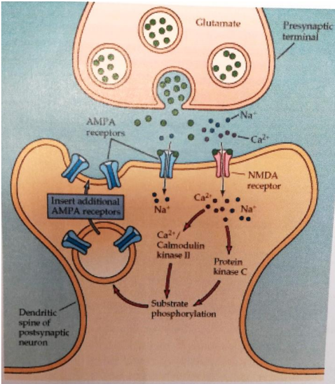
Memory results from a change and a remodeling of connections between neurons involved in a memory system: this is the synaptic plasticity. One of the main mechanisms involved in memory is the long-term potentiation (LTP): an intense and sustainable increase of the synaptic transmission after the stimulation of an afferent fiber (fiber which is going to a central nervous system) with a stimulus of very high frequency. The LTP involves the release of glutamate from a presynaptic neuron which then fix itself on two types of receptors specific to glutamate on the postsynaptic neuron: AMPA receptors and NMDA receptors. Here is a scheme to explain the precise mechanisms of LTP:
Figure 3.1: mechanisms of the LTP
When an action potential arrives in the presynaptic neuron, the vesicles containing molecules of glutamate release the glutamate in the synaptic cleft. The molecules of glutamate bind to AMPA receptors (inducing the depolarization of the postsynaptic neuron) and NMDA receptors, Na+ and K+ ions enter the postsynaptic neuron through AMPA receptors, and Ca2+ ions enter the postsynaptic neuron through NMDA receptors only if the postsynaptic membrane is enough depolarized (otherwise the NMDA receptors are blocked by Mg2+ ions). The Ca2+ ions in the postsynaptic neuron activate proteins kinase, and this pathway insert additional AMPA receptors on the postsynaptic neuron, allowing the LTP to last weeks or months, but it can also be a fundamental process in learning and data storage.
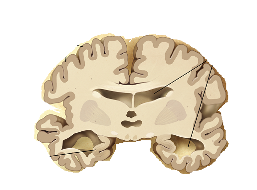
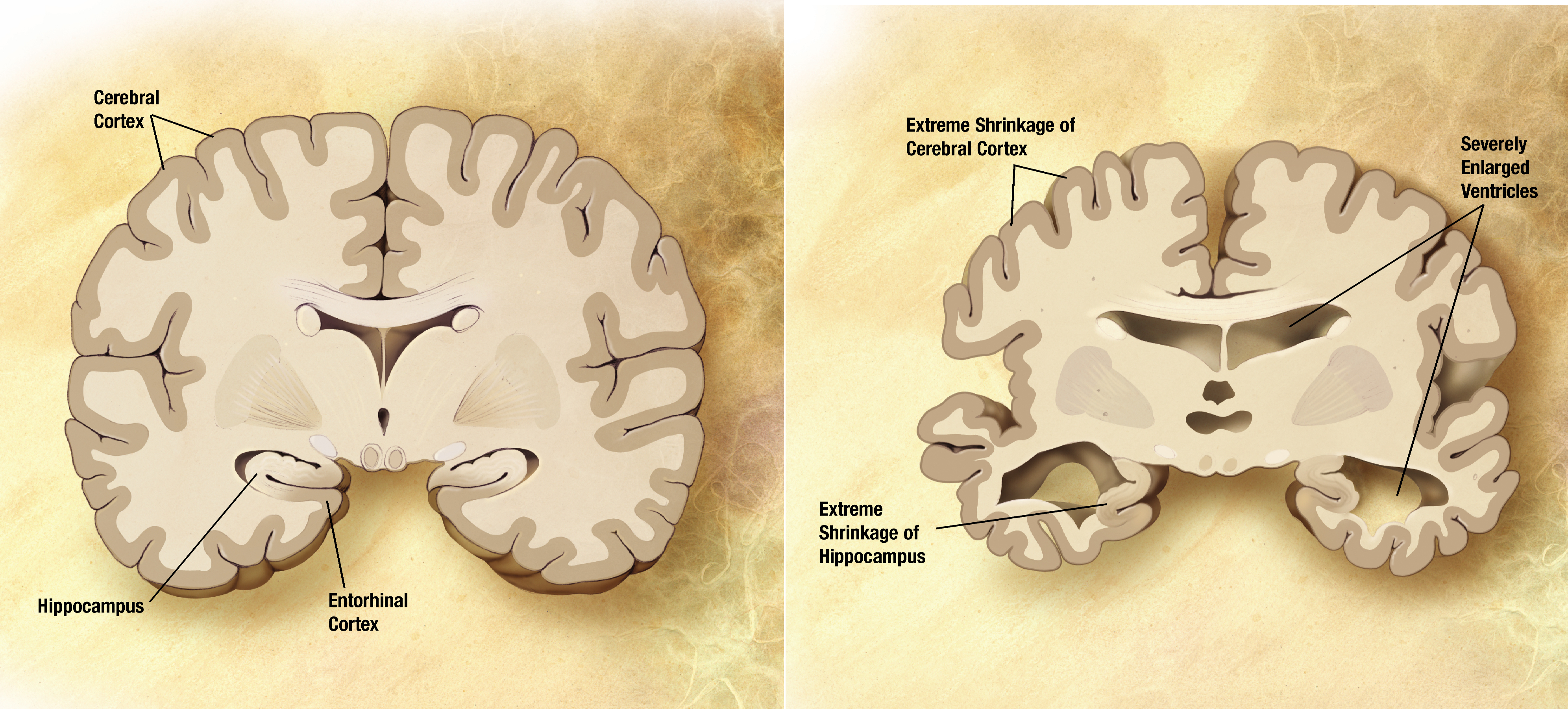
Now, we can talk about Alzheimer’s disease. It causes the death of neurons in the hippocampus and the cerebral cortex, and it involves a neurofibrillary tangle (a neurofibrillary degeneration) and the appearance of extracellular amyloid-beta plaques. Here you can see a normal brain (on the left) versus a sick brain (on the right):
Figure 3.2: brain of a healthy person (left) and brain of an Alzheimer’s disease person (right)
Neurofibrillary tangles are due to an abnormal phosphorylation (hyperphosphorylation) of protein tau and an accumulation of this protein inside the brain tissue. Usually, protein tau binds to microtubules and contributes to the microtubule assembly inside the axons allowing the transport of nutrients. In Alzheimer’s disease, protein tau cannot bind to microtubules, which prevent them to be assembled and they then begin disintegrating. The unbound protein tau forms what we call neurofibrillary tangles. Then, beta-amyloid is a “piece” of a bigger protein called amyloid precursor protein (APP) that spreads out from the inside of the neuron to the outside of the neuron. To do its job, APP is cut by other proteins into several small sections, and sometimes one of the pieces produced is beta-amyloid. In Alzheimer’s disease we can observe plaques of amyloid-beta coming from an accumulation of amyloid-beta that formed clumps of beta sheets leading to the plaques. These plaques collect between neurons and disrupt the cells function.
One final hypothesis about the origin of Alzheimer’s disease has been published in january 2019 in Science Advances by Stephen S. Dominy, Casey Lynch, Florian Ermini et al. Their publication suggests that the bacteria Porphyromonas gingivalis and its toxic proteases called gingipains that have been found in the brain of Alzheimer’s disease patients could be involved in the disease. They infected mice with P. gingivalis and they noticed an increased production of beta-amyloid 42 (a component of beta-amyloid plaques). Also they noticed that gingipains acted as neurotoxins affecting protein tau. They then designed and synthesized a molecule inhibitor targeting gingipains : it blocked the beta-amyloid 42 production, reduced levels of P. gingivalis, reduced neuroinflammation and rescued neurons in the hippocampus.
If you want to learn more about this bacteria involved in Alzheimer’s disease, click here to read the whole article.
2) Parkinson disease
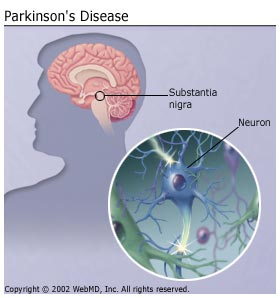
Parkinson disease (PD) is a progressive neurodegenerative disease. A progressive disease evolves over time (becomes worse with time). A neurodegenerative disease means that during the disease there is a loss (or degeneration) in a particular kind of neuron in brain; in this case these neurons are called dopamine neurons. Dopamine neurons make signaling chemicals called dopamine which is essential to make normal movements (functions of dopamine in brain part 2.C). A lot of these neurons are located in the brain, above the spinal cord and midbrain, in a region called substantia nigra (figure 3.3).
These neurons make a huge amount of brain dopamine. When dopamine neurons are lost, by neurodegeneration, there is a reduction in the amount of dopamine in the brain: when we observe this, it is a sign of Parkinson disease. There are 4 main movement signs: 1. Shakiness: often in the form of tremor in hand or fingers. 2. Stiffness: mostly observed when the person tries to bend its body (arm, leg) ,the movement is not enough smooth and fluid, instead they feel rigid. 3. Slowed down movements: it takes much longer for the person to complete the movement, for example compare the speed when you are running in the water or on ground. 4. Balance problems: the person feels really unsteady, unstable when standing or walking around. Not all the patients with PD will have all of these symptoms, but they mostly appear at some point during the disease. Balance problems usually come later, when the person has dealt with the disease after a few years. The first three symptoms usually help in order to detect Parkinson disease. All these main movements signs together form a symptom complex called Parkinsonism or Parkinsonian syndrome. Parkinson is actually the disease that causes Parkinsonism, there are also a few neurodegenerative disease that can cause parkinsonism. Parkinson is the main cause of this syndrome and we often call parkinsonism, Primary Parkinsonism. When the syndrome is caused by an other disease, it is called Secondary Parkinsonism.
The parkinson disease doesn’t only causes parkinsonism. Remember that this a brain disorder and when neurons breakdown there is often more than only one symptom. Patients with PD can also have psychiatric problems such as: – Depression – Cognitive problems such as memory loss – Problem with concentration – Some non movement problems such as smell and problems with their sleeping patterns
The vast part of people having this disease have an idiopathic case, that means the cause is unknown. In the majority of cases there is no family history, but around 15% of cases have family history. In these patients, Parkinson appears to be a subsequence of mutation in a gene (genetic risk factor). Depending on which gene has mutated it can cause (definitely develop the disease) or increase the person’s chance (more likely to do so) to have parkinson disease.
There are also some factors that can increase the chance of developing the disease called risk factors (non-genetic risk factors): – Some pesticides: cleaning chemicals – Age : after reaching 60 years or above – History of Concussions – Gender : Men are a bit more likely to develop this disease (you can read this article for more informations) – Breathing in heavy metals : as copper, manganese Pay attention that this disease doesn’t only happens for older people. About 5-10% of patients are diagnosed before the age of 50 and they are known as “young” or « early onset » parkinson disease (mostly caused by gene mutation).
The treatments of this disease are mostly medications used for minimizing the movements symptoms; these medications increase or replace the dopamine molecule. In most patients the medications works; however, in some patients, when they are dealing with the disease for several years, medication doesn’t really have effects. Another treatment is surgery. The goal of surgery is to inactivate the part of the brain which causes movements problems like shakiness or stiffness that we saw earlier. The biggest issue with this disease is that it is progressive so there is no treatment to really stop the disease.
3) From toxins : botulism, tetanus
We will now speak about neurologic diseases affected by bacterial toxins. In our case botulism and tetanus, these two disease having the same mechanisms, but opposite effects.
Botulism is a disease caused by a toxin created by the bacteria Clostridium botulinum. This toxin blocks the neurotransmission at a neuromuscular junction. The main symptom caused by botulism is known as flaccid paralysis which occurs when muscles are locked in a relaxation state (that can occur if neurotransmitters that are responsible for muscular contraction are blocked for example). In the botulism case, the toxin will inhibit the fusion between vesicles that contains acetylcholine (as we already seen in part 2.A, a neurotransmitter that activate muscular contraction and the muscular membrane) and the membrane of the presynaptic neuron. This fusion occurs thanks to a complex of transmembrane proteins called SNARE. In our case, for the vesicle fusion to occur, two SNARE proteins need to be in contact: synaptobrevin which is located in the vesicle membrane, and syntaxin which is located in the muscular membrane. The contact between SNAREs proteins will lead to the hiring of another muscular membrane protein called SNAP-25 that will bring the vesicle really close to the muscular membrane and so the fusion will begin.
However, the botulism toxin linkage with a receptor usually found in the muscular junction will end up creating an endocytosis vesicle with chemical enzyme in it that will clive a part of the toxin (that become activated when it is clived). It will finally leave the vesicle to go into the muscular junction to cleave a part of SNAP-25 (deactivating it). Without SNAP-25, vesicles will not be able to dock and fuse with the muscular membrane and so muscle contraction by acetylcholine is inhibited leading to a paralysis.
Figure 3.4: Neuromuscular junction and acetylcholine
Note that the botulism toxin is also used to create local muscular relaxation and is commercialized as botox, or onabotulinumtoxinA and used for three main purposes: muscle spasm control, severe underarm sweating and cosmetic improvement.
Tetanus is a disease caused by a toxin created by the bacteria Clostridium tetani. This toxin inhibits the release of inhibitory neurotransmitters into the muscular junction and will thus lead to a uncontrollable permanent state of muscular contraction that is called spastic paralysis. The toxin created by the bacteria is called tetanospasmin and is capable to bind itself with receptors at the end of motoneurons, leading to the internalisation of these toxin by the neuron. Tetanospasmin is then transported into the cell body were it is disposed outside the cell and where it binds to receptors on inhibitory neurons. From there, the toxin enter in a vesicle by endocytosis and, exactly like for the botulism toxin, the acidic pH of the vesicle activates enzymes that cleave tetanospasmin in two sub-units (one used by the other as a channel to escape the vesicle). One of the sub-unit cleaves and inactivates one of the SNAREs proteins called VAMP that is located on vesicles membrane and, without this protein, vesicles containing inhibitory neurotransmitters are not able to fuse with the membrane: muscles will stay in a contracted stated (as excitatory signals can’t be inhibited).
o Text — Alzheimer’s Disease Fact Sheet. (n.d.). Retrieved February 4, 2019, from https://www.nia.nih.gov/health/alzheimers-disease-fact-sheet — C. O. Loubon, J. C. Franco (2010). Neurofisiologia del aprendizaje y la memoria, plasticidad neuronal. iMedPub Journals. Volume 6. No 1:2. — Kocahan S, Doğan Z. Mechanisms of Alzheimer’s Disease Pathogenesis and Prevention: The Brain, Neural Pathology, N-methyl-D-aspartate Receptors, Tau Protein and Other Risk Factors. Clin Psychopharmacol Neurosci. 2017;15(1):1-8.
.jpg&psig=AOvVaw3yIqVGxommZUXwGLDPaBIf&ust=1550060922059118)


